Bidspirit auction | Pernkopf Atlas – An anatomical
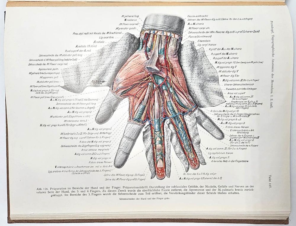
Pernkopf Atlas – An anatomical atlas of the human body illustrated using corpses of Jews sent from the death camps. Berlin–Vienna, 1943 – First edition - Topographische Anatomie des Menschen – The Topographical Anatomy of the Human Body (“The Pernkopf Anatomical Atlas”) by Eduard Pernkopf – published by Urban & Schwarzenberg, Volume I: Part II – Allgemeines, Brust und Brustgliedmaße (General, Thoracic Region, and Upper Limbs). Berlin, Vienna, 1943 – First edition. An anatomical atlas of the human body, illustrated based on the corpses of Jews sent from the death camps to the laboratory of the Nazi anatomist Dr. Eduard Pernkopf, and drawn by a group of artists – one of the most controversial publications of the 20th century. Eduard Pernkopf (1888–1955) was an Austrian anatomist, professor, and dean of the Faculty of Medicine at the University of Vienna during the Nazi period, and a member of the Nazi Party since 1933. During the Nazi era, he served as head of the anatomical institute at the University of Vienna. His colleagues described him as a zealous National Socialist, and he acted accordingly as early as 1938, arriving at work dressed in the uniforms favored by Adolf Hitler. The anatomy department that Pernkopf headed had a history of antisemitism and radical political activity among both students and academic staff, even before Austria’s annexation to the German Reich. On 18 February 1939, the Nazi regime issued an order requiring that all corpses of executed prisoners be sent to the anatomy departments at the nearest universities for research and educational purposes. Later, in 1942, Nazi law ruled that the remains of executed Poles and Jews would not be buried by their relatives, but instead transferred to German research institutions. Many prestigious medical institutions in Germany preserved for research the remains of people who had been murdered. Pernkopf capitalized on the law, working up to 18 hours a day, dissecting bodies in front of teams of artists and illustrators. The shipments did not cease, and the university rooms filled with corpses. “At least half of the 800 illustrations in the atlas were based on the bodies of political prisoners, ” estimated Dr. Sabine Hildebrandt of Harvard Medical School, “they included homosexuals and lesbians, Roma, ideological dissidents, and of course, Jews.” Pernkopf was the man destined to carry out this horrific task, as from the early 1930s, his rapid academic advancement at the University of Vienna went hand in hand with his growing involvement in the Nazi regime. In the same year he began directing the anatomical institute (1933), he joined the Nazi Party, and a year later, the SA (Sturmabteilung). Together with Pernkopf, many of his assistants, technicians, and illustrators who worked with him also joined the Nazi Party. (As dean, Pernkopf insisted on documenting the racial background—Aryan or otherwise—of all faculty members, and demanded that they swear an oath of loyalty to Hitler. He handed over to the authorities the names of those who did not comply with his demands, and in March 1938, all Jews, including Nobel Prize laureates, were expelled from the University of Vienna. Altogether, 132 out of 197 faculty members were dismissed.) By defining certain people as “subhuman, ” Nazi medical science enabled their treatment as “research material” even while they were still alive, stripped of any protection under German law. For years, the book has been regarded as an unparalleled scientific masterpiece, considered one of the most beautiful and precise anatomical atlases ever produced. Pernkopf’s Atlas was described in 1990 by the leading medical journal The New England Journal of Medicine as a “classic work in the field.” It contains hundreds of exceptionally detailed color illustrations, created by artists such as Erich Lepier (who often signed his name alongside a swastika), Karl Endtresser, Ludwig Schrott (all three were Nazi activists), and others. Each illustration was drawn by hand, at full scale, with astounding precision. Rich in detail, the atlas provides uniquely sharp and graphic depictions of skin and bone, the muscular and tendon systems, nerves, and human organs. The illustrations in the book have been used—for decades—by physicians, surgeons, and medical students, and even today, in certain contexts, the atlas is still in use. Leading anatomists in the medical world have testified over the years: “You cannot even begin to compare it to other books in terms of precision and attention to detail.” Doctors worldwide have stated that during complex surgeries, the book helps them identify which of the many nerves operating in the body is the true source of pain—more than any other diagnostic tool available to this day! In the years immediately following the war, the atlas was used in good faith by physicians in several countries, without awareness of the origins of its illustrations. Over time, however,